Does eating marinated meat cause cancer?
How to optimize the SpectraMax Paradigm multi-well microplate detector for Transcreener fluorescence polarization detection test
Introduction
This article describes how to optimize instrument parameters to meet validation requirements when using the SpectraMax ® Paradigm® Microplate Detector from Molecular Devices to detect Bellbrook's Transcreener Fluorescence Polarization Kit.
• Transcreener ADP2 FP (3010)
• Transcreener AMP/GMP (3006)
• Transcreener UDP FP (3007)
• Transcreener GDP FP (3009)
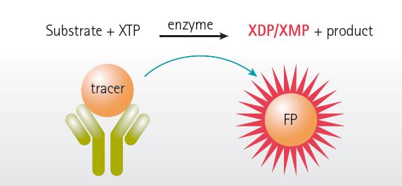
The Transcreener high-throughput screening test is based on a versatile, high-throughput biochemical detection platform that detects the ADP content of thousands of different cellular enzymes in the system after catalyzing the substrate. Most of these enzymes catalyze covalent reactions in cellular signaling pathways and are high value targets for drug screening. Transcreener fluorescence polarization test uses a single-step, immuno-competition method to directly detect the nucleotide polarization values ​​of far-infrared fluorescent tracer molecules. All the detection reagents are from far-infrared fluorescent tracer molecules and highly specific monoclonals. Or a combination of polyclonal antibodies, the principle is that after the target enzyme catalyzes the substrate, the produced nucleoside diphosphate or single nucleotide competitively replaces the far-infrared fluorescent tracer molecule linked by the antibody, and its far-infrared fluorescence shows The rotational freedom of the tracer is increased, resulting in a weakening of the fluorescence polarization phenomenon (Fig. 1). The use of far-infrared fluorescent tracer molecules can effectively reduce the interference of fluorescent compounds and light scattering. Transcreener fluorescence polarization tests are added in one step, and mixed and read. The way is especially suitable for high-throughput screening.
Transcreener fluorescence polarization test principle (Figure 1)

The SpectraMax Paradigm Multi-Purpose Plate Detector is designed with a flexible cartridge design that allows you to select the most suitable test cartridge for different test lab types for optimal results. The instrument has dual photomultiplier diodes (PMT) that can work simultaneously. It is used to detect the fluorescence polarization signal value, so as to improve the detection speed without reducing the detection sensitivity. The instrument not only supports 1536-well plates, but also integrates with the company's StakMax® stacker, which greatly increases the speed and throughput of inspections. For the instrument settings to optimize the fluorescence polarization characteristics Transcreener test, SpectraMax Paradigm Multifunctional Microplate Reader performance well beyond the requirements of the test instrument performance itself.
Verification standard
In order to take advantage of Transcreener's high-throughput screening test, the key factor is how to set and optimize the parameters of the microplate detector. For the type of test, the correct parameter setting and optimization will increase. The sensitivity of its detection. To find the optimal parameter settings for the Transcreener high-throughput screening test, we made 24 replicates of a standard curve simulating the reaction of the 10uM ATP/ADP enzyme. The initial concentration is 10uM ATP, and the increase of ADP content is proportional to the decrease of ATP content, and the total adenine nucleotide concentration is maintained at about 10uM. By changing the different detection time (Integration time) to meet the requirements of Test 0.5 Z value>, verify that an instrument can meet the requirements Transcreener fluorescence polarization tests, in which 10% of the Z value 10uM ATP conversion rate of> 0.7 and ΔmP > 120.
material
• ATP/ADP mixture - 4 mM MgCl 2 , 2 mM EGTA, 50 mM HEPES, pH 7.5, 1% DMSO
0.01% Brij-35 and ATP/ADP (adenine concentration 10uM)
• ADP Detection Mixture - 1x Termination & Detection Buffer B, 4 nM ADP Alexa633 Tracer and 14.8 ug/ml ADP 2 Antibody
• Single Tracer-1x Stop Solution & Detection Buffer B and 4 nM ADP Alexa633 Tracer
• Blank Buffer-1x Stop Solution & Assay Buffer B and 14.8 ug/ml ADP 2 Antibody
Detailed testing steps, including preparation of the standard, please refer to the Transcreener Technical Manual (http://)
method
Test preparation
Step 1: Add 10 ul of ATP/ADP compound to each row of the 384-well plate
Step 2: Add 10ul of ADP to these rows and mix
Step 3: Add 10 ul of 10 μM ATP/0 μM ADP compound to row P in a 384-well plate.
Step 4: Add 10 ul of single tracer molecule to each well in the P1-P12 row
Step 5: Add 10 ul of blank buffer to each well of the P13-P24 row.
Instrument settings
Table recommended microplate reader SpectraMax Paradigm provided | |
parameter | Setting |
Test cartridge | Fluorescent polarization card box |
Filter: Wavelength / Bandwidth | Excitation light: 624/40 nm; emission: 684/24 nm |
Detection type | End point method |
PMT and light path | Set the detection time of 20 milliseconds or longer in Stop and Go mode. Or use on-the-fly mode |
Reading height | When using a Corning 3676 microplate for 6.61 mm |
The SpectraMax Paradigm Microplate Detector is controlled by SoftMax® Pro software, which allows you to select a template file for this test type and change its specific parameter settings in its software template library.
Step 1: Open the SoftMax Pro operating software, click the 'Protocol Manager ' option, select the pre-set template in its 'Protocol Library' template library, and select the 'FP Rhodamine' template in the 'Paradigm Protocol ' folder.
Step 2: Click 'Plate01' in the navigation tree on the left side of the software, then click 'Settings' on the right side to set the button (gear icon), the setting dialog box appears.
Step 3: Select Alexa Fluor633 Fluorescence Polarization Cartridge
Step 4: The detection mode selects 'FP', that is, the fluorescence polarization mode, and the detection type selects 'Endpoint', that is, the end point method.
Step 5: The card box has its detection wavelength fixed, no need to set it again.
Corning low vol/rndbtm'
Step 7: Click on 'Read Area ' to select the microplate detection area (hole)
Step 8: Click on 'PMT and Optics' to select 'Off – Stop and Go ' or 'Performance – On the fly' to read the speed mode.
Step 9: If you select Stop and Go mode, enter a value of 20ms or higher at 'integration time ' to extend the time for better results, but will increase the detection time.
Step 10: If the best detection height is known, input it directly. When using the new test or new microplate, the height optimization should be re-set. The original value before height optimization is 1mm. After optimization, the new height is optimized. The value will replace the original value
Step 12: Select 'Show Pre-Read Optimization Options'
Step 13: Click the 'OK ' button to close the settings dialog
Step 14: Click the 'Read' button, an optimization selection dialog box will appear in the center of the screen, and the microplate optimization and detection height optimization should be performed again for the new test.
result
Sample fluorescence polarization standard curve
During the reaction, as the ADP content increases, its ratio to ATP changes, and the tracer molecule bound to the anti-ADP antibody decreases, resulting in a decrease in the fluorescence polarization signal value in the reaction system. SpectraMax Paradigm is given for this test. After 15 data points were detected, the values ​​were automatically fitted to generate a standard curve.
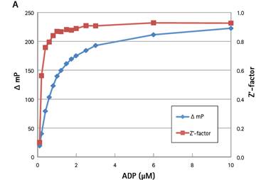
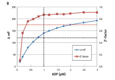
Test verification parameters (Figure 2 )


A: Simulate the Z value and Δ mP value of the standard curve when converting 10uM ATP to ADP ; B: When the microplate detector is set to 100ms detection time, the standard curve view is enlarged, and the ADP concentration range is displayed between 0-3uM. Verify the minimum data point for the Z value (red dashed line) and the minimum data point for verifying the Δ mP value (horizontal black dotted line), also showing the 10% ATP conversion verification point (vertical black dotted line)
Table II exhibit different detection results provided by | |||||||
10uM ATP conversion rate is 10% | |||||||
Detection time | 20ms | 30ms | 50ms | 100ms | 150ms | 200ms | OTF |
Reading time | 1:02 | 1:08 | 1:17 | 1:42 | 1:59 | 2:17 | 0:55 |
Δ mP value at 0% ATP conversion | 147 | 146 | 144 | 139 | 136 | 151 | 148 |
Z value at 10% ATP conversion rate | 0.77 | 0.80 | 0.79 | 0.87 | 0.84 | 0.91 | 0.77 |
in conclusion
Acknowledgement
Molecular Devices expressed her heartfelt gratitude to Ms. Meera Kumar of Bellbrook Laboratories for their great cooperation.
Cesarean Section Surgical Pack
Cesarean Section Kit,C-Section Drape Pack,Cesarean Section Drape Set,Cesarean Section Surgical Drape Pack
Suzhou JaneE Medical Technology Co., Ltd. , https://www.janeemedical.com