Keywords: soluble antigen soluble antigen solubleantigen glycoprotein lipoprotein cell homogenate
Proteins, glycoproteins, lipoproteins, enzymes, complements, lipopolysaccharides, bacterial exotoxins and nucleic acids are all soluble antigens, and a considerable part of them are derived from tissues and cells, and the composition is complex. In the preparation of such immunogens, the tissue and cells must first be disrupted, and then the target protein or other antigen is extracted from the tissue and cell homogenate, and the purified antigen needs to be identified before being used as an immunogen.
First, the preparation of tissue homogenate
The material used to prepare the immunogen must be fresh or cryopreserved. Immediately after the material is obtained, the capsule or connective tissue is removed. The organ should be irrigated to remove the residual blood in the blood vessel. The blood and dirt are washed away with physiological saline containing 0.5 g/L NaN 3 ; in a 4 ° C water bath or ice bath. The tissue to be washed was cut into 0.3-0.5 cm3 pieces, and an appropriate amount of physiological saline was added thereto, and the tissue homogenate was prepared by using a tissue grinder (OMNIBead Ruptor 24 multi-sample grinding bead homogenizer). After the tissue homogenate was centrifuged at 3000 r/min for 10 min, the supernatant was used as a material for extracting soluble antigen. The supernatant must also be centrifuged to remove cell debris and minute tissue prior to extraction.
Second, cell breakage
To extract the soluble antigen of the cell, the cell needs to be broken. Depending on the type of cell, there are some differences in the method of selecting the broken one. Several commonly used methods of cell disruption are introduced.
1 , enzyme treatment
Lysozyme, cellulase, snail enzyme, etc. can digest bacteria and tissue cells under certain conditions. For example, lysozyme has a bacteriostatic effect on the cell wall of Gram-positive bacteria. The enzyme treatment method is suitable for the dissolution of a plurality of microbial cells, and the method has the characteristics of mild action conditions, unbreakable inclusion components, and controllable cell wall damage.
2 , freezing and thawing method
The cells are mainly destroyed by sudden freezing, formation of intracellular ice crystals, and sudden changes in the concentration of intracellular and extracellular solvents. The method is to completely freeze the broken cells in a refrigerator at -15 to -20 ° C, and then take them out of the refrigerator and let them slowly melt at 30 to 37 ° C. This is repeated twice, and most of the tissue cells and particles in the cells can be broken. This method is applicable to tissue cells and has a poor effect on the cells of microorganisms.
3 , ultrasonic crushing method
This is a method of breaking cells by mechanical vibration of ultrasonic waves. Due to the cavitation during the occurrence of ultrasonic waves, the liquid is partially decompressed, causing internal flow of the liquid, and when the vortex is generated and disappears, a large pressure is generated to break the cells. The frequencies used for ultrasonic waves vary from 1 to 20 kHz. When performing ultrasonication, it needs to be carried out intermittently to avoid prolonged ultrasonic heat production, resulting in antigen destruction. Ultrasonic pulverized cells can also be placed in an ice bath to cool down. Ultrasonic disruption of cells is simple, reproducible, and saves time. This method can be used for the disruption of microorganisms and tissue cells. Optional ultrasonic cell disrupter (OMNI Sonic Ruptor400W)
4 , surfactant treatment
At appropriate temperatures, pH, and low ionic strength, the surfactant can form microvesicles with lipoproteins, which lyse the cells through changes in cell membrane permeability. Commonly used surfactants are sodium lauryl sulfonate (SDS cationic type), chlorhexidine, Triton X-100 and the like. For example, 1 g of Escherichia coli wet cells can be lysed by adding 2% Triton X-100 for 60 min. This method works mildly. This method is commonly used to break up cells when extracting nucleic acids.
Third, the purification of protein
The protein purification method belongs to biochemical technology, and this chapter only gives a brief introduction.
1 , ultracentrifugation
The principle of separating and purifying the antigen by the method is to utilize the difference of the sedimentation speed of each particle in the gradient liquid, so that the particles with different sedimentation speeds are in different density gradient layers to achieve the purpose of separating from each other. Commonly used density gradient media are sucrose, glycerin, CsCl, and the like.
When separating and purifying antigen by ultracentrifugation or gradient density centrifugation, it is extremely difficult to separate an antigen component except for individual components, so it is only used for the separation of a few macromolecular antigens, such as IgM, C1q, thyroglobulin, etc. Some lighter-weight antigenic substances such as apolipoprotein A, B, etc. Most medium and small molecular weight proteins are difficult to purify by this method.
2 , selective precipitation method
The principle is based on the difference in the physical and chemical properties of each protein, using various precipitants or changing certain conditions to promote the precipitation of protein antigen components, thereby achieving the purpose of purification. The most common method is the salting out precipitation method.
Principle of salting out
The solubility of a protein in an aqueous solution depends on the number of water molecules around the surface of the protein molecule, that is, the extent to which the peripheral hydrophilic group of the protein molecule forms a hydrated film with water and the charge of the protein molecule. After the neutral salt is added to the protein solution, since the affinity of the neutral salt to the water molecule is greater than that of the protein, the hydration layer around the protein molecule is weakened or even disappeared. At the same time, when the neutral salt is added to the protein solution, the ionic strength changes, the charge on the surface of the protein is largely neutralized, and the solubility of the protein is lowered, so that the protein molecules aggregate and precipitate. Due to the different solubility of various proteins in different salt concentrations, different saturation salt solutions precipitate proteins that separate them from other proteins. The most commonly used salt solution is ammonium sulfate of 33% to 50% saturation. The salting out method is simple and convenient, and can be used for crude extraction of protein antigens, extraction of gamma globulin, concentration of proteins, and the like. The purity of the antigen purified by salting out is not high, and only the initial purification of the antigen is applied.
3 , gel chromatography
Gel chromatography is the separation of proteins by molecular sieves. The gel is a material having a three-dimensional porous network structure, and after being equilibrated by a suitable solution, it is loaded into a chromatography column. When a sample solution containing various molecules slowly flows through the gel chromatography column, the macromolecular substance does not easily enter the micropores of the gel particles, and can only be distributed between the particles, so the speed of downward movement during elution Faster, first eluted. In addition to being diffused in the interstitial space of the gel particles, the small molecule material can also enter the micropores of the gel particles, and the downward movement rate is slower when eluting, and then eluted. Therefore, protein molecules are separated by molecular size.
4 , ion exchange chromatography
The principle of ion exchange chromatography is to use a number of cellulose or gels with ionic groups to adsorb and exchange oppositely charged protein antigens. Since the isoelectric points of various proteins are different, the amount of charge carried is different, and the ability to bind to cellulose (or gel) is different. When the gradient elutes, the ionic strength of the mobile phase is gradually increased, so that the added ions compete with the protein for the charge position on the cellulose, thereby dissociating the adsorbed protein from the ion exchanger.
The ion exchangers commonly used in ion exchange chromatography have the following
1 cellulose with ion exchange groups, such as carboxymethyl (CM) cellulose, DEAE-cellulose
2 cross-linked dextran, agarose and polyacrylamide with ion exchange groups
3 Gel-synthesized highly crosslinked resin.
5 , affinity chromatography
Affinity chromatography is a chromatographic technique designed to take advantage of the biological specificity of biomacromolecules, that is, the specific affinity between biomacromolecules. For example, antigens and antibodies, enzymes and enzyme inhibitors (or ligands), enzyme proteins and coenzymes, hormones and receptors, IgG and staphylococcal protein A (SPA) have a special affinity. For example, when purifying IgG, the SPA can be adsorbed on an inert solid phase substrate (such as Speharose 2B, 4B, 6B, etc.) and prepared into a chromatography column. When the sample flows through the column, the IgG to be separated can specifically bind to the SPA, and the remaining components cannot bind to it. After the column is sufficiently eluted, the ionic strength or pH of the eluate is changed, and the IgG is dissociated from the SPA on the solid phase substrate, and the eluate is collected to obtain the IgG to be purified.
The main advantage of affinity chromatography for purification of protein antigens is high purity, simple and fast, but more expensive.
Fourth, the preparation of nucleic acid antigen
Nucleic acid molecules are immunogenic and can be used in immunogens to prepare antibodies. The main step in extracting nucleic acids is to first break up the cells to free the nucleic acids from the cells, extract them with phenol and chloroform to remove the proteins, and finally precipitate the nucleic acids with ethanol.
5. Preparation of lipopolysaccharide antigen
Lipopolysaccharide (LPS) is an important component of the cell wall of Gram-positive bacteria and has a variety of biological effects. The LPS is usually extracted by the phenol method. The specific method is: mixing the dried cells (or a relatively large number of wet cells) in water, heating to 65-68 ° C, adding an equal volume of pre-warmed phenol, and stirring vigorously, heating for 5 min, immediately using ice cold The water was rapidly cooled down to below 10 ° C and centrifuged at 3000 rpm for 30 min to separate into two layers. The upper layer is the water layer, the lower layer is the phenol layer, and the bacterial residue is at the bottom. Aspirate the aqueous layer (including LPS), remove the phenol by dialysis, concentrate, and ultracentrifugation. The lipopolysaccharide is located in the transparent gelatinous part of the upper layer, and is taken out in water and centrifuged to obtain a purified LPS sample.
Identification of purified antigen
In order to obtain a good immune effect, the antigen should be identified after purification to be used for animal immunization. The identification of antigen mainly includes the following aspects:
1 , content detection
The most accurate method for determining the protein content is the Kjeldahl method, but it is not suitable for general laboratories because of the requirement for precision equipment. The general laboratory uses spectrophotometric measurements. The method first measures the absorbance at 280 nm and 260 nm (A), and then calculates the protein content using an empirical formula.
Protein content (mg/ml) = A280 x 1.45 - A260 x 0.74.
In addition, the protein content can also be used in the Folin phenol method, the specific operation see clinical biochemical technology.
2 , the identification of molecular weight
The molecular weight is generally determined by SDS-PAGE electrophoresis, as detailed in clinical biochemical techniques.
3 , purity identification
It is often identified by zone electrophoresis. See clinical biochemical technology for details.
4 , identification of immune activity
A two-way agar diffusion test is often used.
This experimental scheme uses Omni's Bead Ruptor 24 multi-sample grinding bead homogenizer and Prep multi-sample homogenizer (provided by Beijing Pingliyang Co., Ltd.).
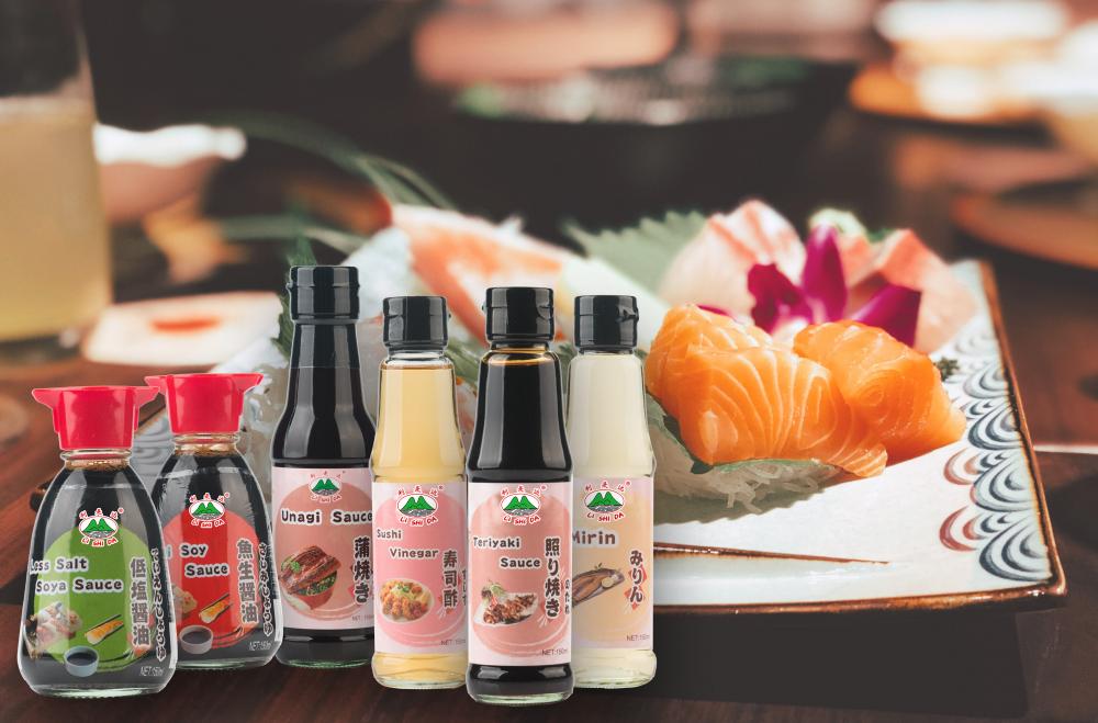
In order to satisfy people's need,now we develop Japanese Sauce,includes Sushi Sauce,Sashimi Sauce,Unagi Sauce,Teriyaki Sauce,Sushi Vinegar, Mirin Sauce and so on.We select non-GMO soybeans,wheat,sugar and other high quality ingredients,and adhere to brew in traditional process.

Welcome to contact us for cooperation!
Japanese Soy Sauce,Sushi Soy Sauce,Sashimi Soy Sauce,Unagi Sauce,Teriyaki Sauce,Sushi Vinegar
KAIPING CITY LISHIDA FLAVOURING&FOOD CO.,LTD , https://www.lishidafood.com