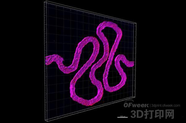
In March of this year, a research team led by Jennifer A. Lewis , a professor of bioengineering at Hansörg Wyss at Harvard University, invented a method that could use human stem cells, extracellular matrices, and circulatory channels lined with vascular endothelial cells. 3D printed a thick vascularized tissue structure and maintained activity for more than a month. Now that the team goes one step further, 3D creatures have printed a tubular 3D kidney structure that reproduces the function of the kidneys and takes a step closer to bioprinting functional human tissues and organs.
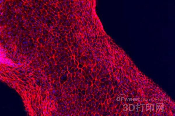
Working closely with Roche Pharmaceuticals scientist Annie Moisan, they have built a functional 3D kidney structure based on the previous structure that contains living human epithelial cells that make up the surface of the renal tubules. The study is currently published online in the journal ScientificReports.
“The current work further extends our bioprinting platform to create a functional human tissue structure that is both technical and clinical,†said Professor Lewis.

According to Xiaobian, the 3D kidney structure created by Lewis's team simulates the proximal tubule, the longest and thickest segment of the tubule, and an important component of each nephron. The so-called nephron is responsible for the key function of switching between blood and urine. Each side of the kidney has 100-1.5 million nephrons. At the curl of the proximal tubules, approximately 65-80% of the nutrients are reabsorbed from the kidney filtrate and transported back to the blood. Therefore, the research team's bioprinted 3D kidney architecture reproduces a very small – but very critical – subunit of the entire kidney.

ZHEJIANG SHENDASIAO MEDICAL INSTRUMENT CO.,LTD. , https://www.sdsmedtools.com